MRI - Neolife Bucharest High Performance Medical Imaging Services
What is the MRI? (Magnetic Resonance Imaging)
Magnetic Resonance Imaging (MRI) is a high-performance imaging method, non-invasive and non-radiant, that uses a strong magnetic field in order to obtain fine details of various components of the human body. By MRI all organs can be investigated, the method being selective from the disorders of the nervous system, genital and muscle and bone systems.
In certain cases (with the exception of patients with advanced kidney failure) the patients can be administered a paramagnetic contrast agent, in order to characterise and delineate the lesion and in order to differentiate it from other pathologies. Allergic reactions to the paramagnetic contrast agent are very rare.
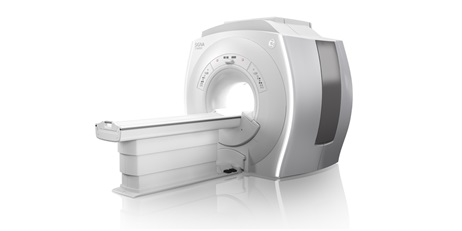
Neolife Medical Center Bucharest completes its imaging department with a high-performance MRI device, Signa Explorer 1,5 T , produced by General Electric
Depending in the investigated region, the examination may take between 20 and 60 minutes, but may be extended depending on the investigated area.
This investigation doesn’t require special patient preparation (pre-examinations, special diets, or medication). The patient shall enter into the examination room without jewelry, watches, bank cards, hearing aids - these can be damaged due to the strong magnetic field or they can interfere with it.
During the MRI investigation, the patient shall lie on a mobile table that is inserted into a tunnel. The patient shall not feel any discomfort, but shall hear certain sounds produced by the device (intermittent or continuous). Throughout the duration of the examination, the patient shall be under the observation of the operator who carries out the investigation and may talk to him/her.
Necessary information for the MRI investigation
Prior to the carrying out of the MRI the Medical Consultant must be informed whether:
Another specific situation is represented by the maternal pathology during pregnancy. Although up to the present moment no adverse effects of this type of investigation have been proven, in the first three months of pregnancy MRI must be avoided - examination can be done only if the benefit is greater than the risk that the fetus is subjected to. If the investigation is nonetheless necessary, the Attending Physician together with the Radiologist shall assess the risk/benefit report and shall decide the opportunity of the examination.
Manner of carrying out the MRI investigation
The MRI investigation shall be carried out by the Radiologist.
The patient shall remove from his/her body all metal objects (hearing devices, dental plaque, any types of jewelry, watch and hairpins) as there is the risk that these objects might be attracted by the magnet used for the carrying out of the examination.
The patient must be completely undressed depending on the area examined (in certain cases, patients may keep part of the clothes, if they are not an inconvenience). The patient may use the disposable robe supplied by the clinic. If certain clothes may be kept, the patient must empty his/her pockets of coins, telephones or cards (for instance, credit cards or ATM cards) with magnetic bands inscribed on them, as by the MRI investigation they can demagnitize).
During the examination, the patient shall lie on the back on the table of the device, which represents the device scanner. The head, thorax and limbs must be fixed with certain devices (antennas) in order to keep the patient still. The table shall slide inside the device that contains the magnet. A device in the shape of a ring (antenna) may be placed over or around the region that is about to be scanned.
Inside the scanner, the patient shall hear a ventilator and shall feel the air moving. Moreover, he/she can also hear various noises that are the result of the scanning. The device is equipped with headphones and earplugs in order to reduce the noise.
It is very important that the patient does not move during scanning. Furthermore, the patient shall be asked to hold his/her breath for short periods of time. During the carrying out of the test, the patient shall be locked into the scanning room, but the medical consultant shall monitor the patient by means of a transparent window. The patient shall communicate through a microphone with the physician carrying out the investigation.
If the use of a contrast agent is necessary, it shall be administered at the level of peripheral veins of the patient’s arm. The contrast agent shall be administered in 1 to 2 minutes.
In rare cases, the following may emerge:
TO BE REMEMBERED! - The MRI investigation is a complex and painless investigation.
Releasing the result
The written result shall be released within 48-72 hours as of the moment of the carrying out of the investigation.
The result shall be released based on an identification document.
Information for the scheduling of an investigation and what documents are necessary:
Appointments shall be made on the telephone number: 021 9230 only if you have a written recommendation from a physician.
Documents needed:
Advantages of Magnetic Resonance
The short time of image acquisition due to the gradients available and the top of the range antennas
High-resolution images by the use of surface antennas for all body parts, especially for joints.
Applications of MRI investigation
Morphological and functional efficient evaluation by:
Evaluation by sequences dedicated to incipient lesions of the articular cartilage and aiming at its recovery after the intraarticularly administered treatments.
Evaluation of micro-haemorrhages and of the vein components of cerebral blood malformations by gradient sequences carried out with very thin sections.
Colon examination with the highlighting of the lumen and walls with the evaluation of parietal and extraparietal extension of potential pathological processes simultaneously with the scanning of the liver for concomitant lesions.
Examination of the urinary system with RM urography which offers images similar to the classic urography without and with the injection of paramagnetic contrast agent simultaneously with the evaluation of the vascularization and the renal function through the parenchymatous loading and elimination of the agent contrast through the urinary system.
MRI Medical Team - Neolife Bucharest
 Dr. Mirvald Maro
 Dr. Oprisescu Bogdan
 Dr. Teodorescu Bogdan